UVA Labs Map the Part of Our Brain That Helps Us Select Our Path

The labs of University of Virginia professors Jianhua “JC” Cang and John N. Campbell have created the first comprehensive molecular atlas of the important brain region called the superior colliculus, which helps us direct our gaze toward objects and scenes of interest as we decide how to move through the world.
The new study, led by graduate student Yuanming Liu and postdoctoral fellow Elise Savier, appeared online Friday in the journal Neuron.
“Direction selectivity is a fascinating feature of the visual system that’s been studied for over 60 years, though the genetic basis for direction selectivity has been unclear,” said Campbell, an assistant professor of biology.

Jianhua “JC” Cang is an expert in mammalian visual systems. (Photo by Dan Addision, University Communications)
The superior colliculus can “see” without “seeing.” By monitoring the shift of our gaze, this area of the midbrain, located above the brainstem, contributes to our mental map and helps us determine what needs attention. It processes visual stimuli independent of the visual cortex and in coordination with other sensory information.
Patients whose primary visual cortex is damaged can sometimes point to visual objects they say they can’t see. The phenomenon is called “blindsight.” This happens because the superior colliculus compensates.
Putting Their Minds Together
Cang, Paul T. Jones Jefferson Scholars Foundation Professor of Neuroscience and director of the undergraduate Program in Fundamental Neuroscience, is an expert in mammalian visual systems. He said the team used mice as the research model because their visual pathways are similar to our own.

John N. Campbell’s lab examined the genetic profile of the superior colliculus. (Biology Department photo)
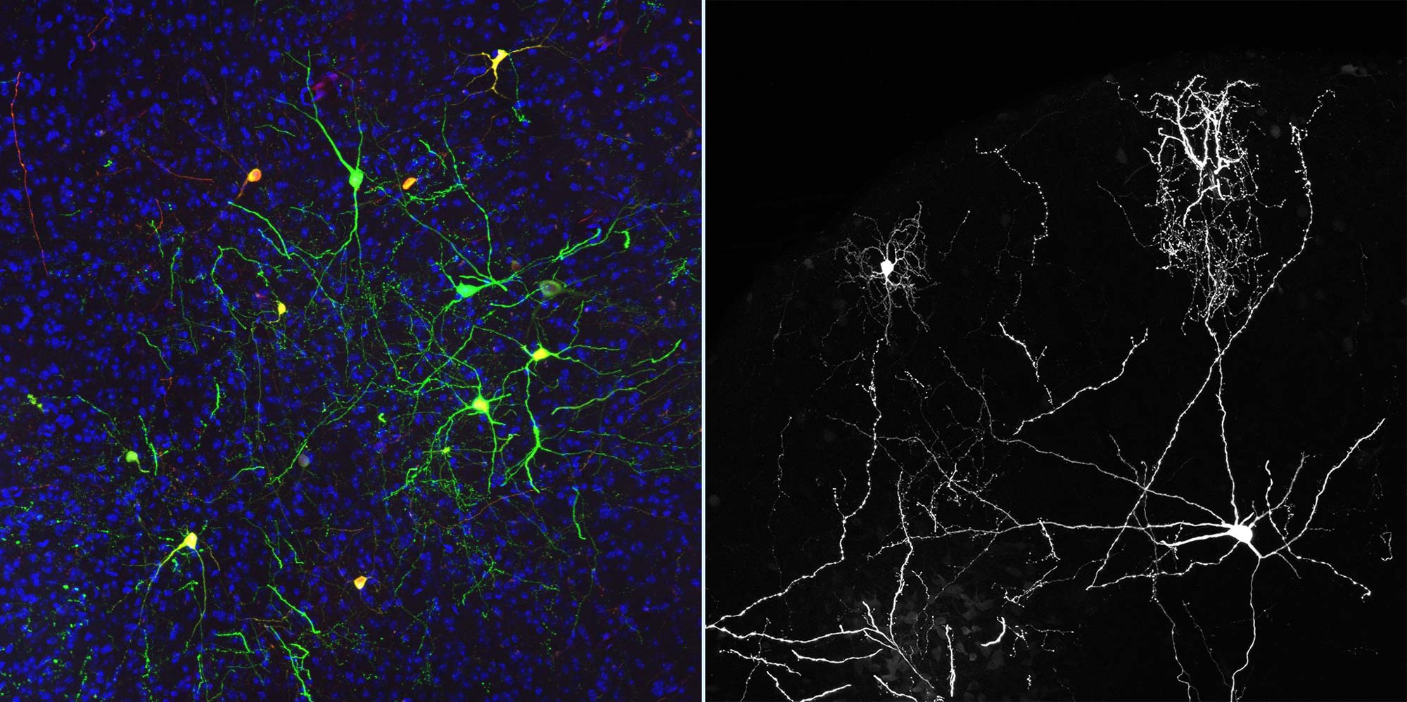
Specifically, his lab studied how individual neurons in the brain, including those in the superior colliculus, processed an assortment of visual stimuli. This typically involved black-and-white bars on a computer screen that drifted in various directions in front of the mice, which were under a microscope. The researchers were able to see what was going on in the mice brains through what’s known as “two-photon calcium imaging.” The technique detects calcium, which increases with neuronal activity, by using fluorescent imaging.
Campbell’s lab then examined the genetic profile of the same neurons.
“Our labs teamed up to develop a method in which we could image a single neuron’s activity in awake, behaving mice, then identify that same neuron afterward based on the genes it expresses,” Campbell said. “In the process, we generated a comprehensive catalog of the molecular cell types that make up the superior colliculus, including many not previously characterized.”
Mapping the Brain – A Grand Challenge
The research suggests that the cell types identified may play distinct roles in visual processing. This study lays the foundation for future research.
The latest findings are in keeping with the UVA Grand Challenge to expand the boundaries of neuroscience with research that maps the brain and helps pioneer the way toward life-improving health advances.

These microscope images demonstrate two ways researchers were able to view neuronal change. (Images by Elise Savier)
“Given superior colliculus’ importance in vision and attention, including in such disorders as blindsight and spatial neglect, as well as in decision-making, which is integral to neurological disorders of cognition, our research will lead to a better understanding of all these conditions,” Cang said.


